


Department of Visual Electro Physiology
Visual Electro Physiology
 Occasionally a person experiences visual changes that are difficult to completely understand with only a clinical eye exam. These changes may be due to abnormalities in the function of some of the cells in the retina, or there may be changes in the nerve connections between the eye and the brain. Sometimes it is difficult to find out the exact point of damage in the pathway of vision or to determine how well babies can see.
Electrophysiology testing includes a battery of tests which can be used to provide information about the visual system beyond the standard clinical examination of the eye. Each image we see is interpreted to an electrical sign. It is possible to detect this electrical response each time a light flashes or a stimulus moves or suddenly appears. This response comes either from retina, the light-detecting part of the eye (electroretinogram, ERG) or the region of the brain that is responsible for vision (visual evoked potentials, VEP). We have the ability to detect these responses through electrodes placed at specific parts
Tests offered
- Electroretinogram (ERG) – Full field ERG, Pattern ERG. An ERG is useful in evaluating inherited (hereditary) and acquired disorders of the retina such as retinitis pigmentosa, Leber’s congenital amaurosis, cone dystrophies, etc. It is often ordered if there is a complaint of night blindness.
- Multifocal ERG (MFERG) – Ordered in macular disorders
- Visual Evoked Potentials (VEP) – Flash VEP, Pattern VEP. VEP tests are used to evaluate optic neuritis, optic tumors, demyelinating diseases such as multiple sclerosis and intracranial lesions, including brain tumors
- Electro-oculogram (EOG) – Ordered in inherited retinal dystrophies.
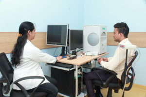
Procedure
For ERG, a thin wire with an electrode is placed on the eye after topical anesthesia, while for VEP electrodes are attached to the head. The patient then watches a screen with different lights to stimulate the retina. The electrical impulses from the retina are recorded. To get useful readings, the patient must first sit in the dark for a period of time to become completely dark-adapted. For this reason, this test usually takes about two hours.
ICARE Eye hospital is one of the few eye care centers that provide a full complement of electrophysiological tests for visual function assessment in the diagnosis, prognosis, and treatment of eye diseases. These examinations are held daily in our Institute by trained staff, they are not at all invasive and they are completely safe for babies and infants as well.
Related Consultants
-

Dr. Geetha Srinivasan
- Senior Consultant, Pediatric Ophthalmology and Strabismus
- MBBS, MS, DNB
- drgeetha@icarehospital.org
-

Dr. Kanika Gupta
- Consultant - Pediatric Ophthalmology
- MBBS, FRCS(Glasgow) part-1, MS(Ophthalmology), FICO(advanced), DNB(Ophthalmology), Fellowship(Pediatric Ophthalmology)
- drkanika@icarehospital.org